

Portable Ultrasound for Veterinary Ophthalmology
Hand-held Fundus Camera
FUNDUS CAMERA
WIRELESS ULTRASOUND
Hand-held Fundus Camera
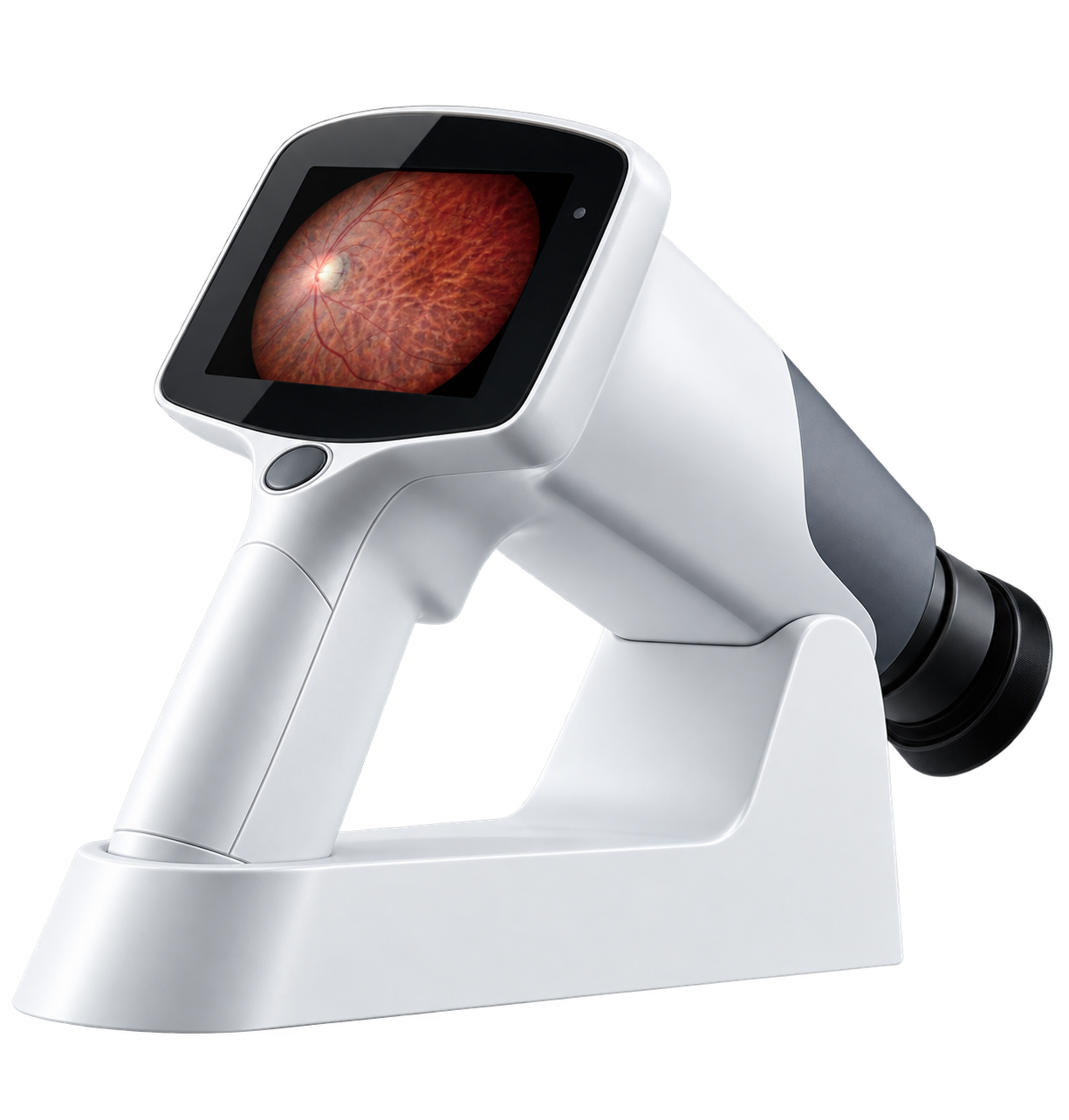
Capture detailed retinal images to support documentation, diagnosis, monitoring, and client communication with veterinary-focused fundus imaging.
Capture detailed retinal images to support documentation, diagnosis, monitoring, and client communication with veterinary-focused fundus imaging.

See the FC161 in Action
Watch a 3-minute overview of the FC161 Hand-held Fundus Camera and see how easily it captures high-quality retinal images in a handheld workflow.
Watch a 3-minute overview of the FC161 Hand-held Fundus Camera and see how easily it captures high-quality retinal images in a handheld workflow.
ADVANCED TECHNOLOGY
Auto Split Focusing Technology
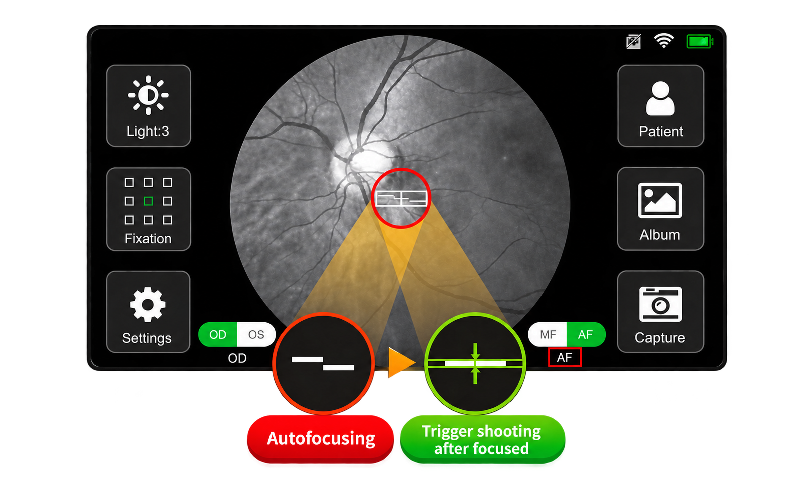
The FC161 uses Auto Split Focusing technology with a focusing speed that reaches milliseconds. It helps reduce out-of-focus images caused by patient movement, improves image quality, and reduces the need for cooperation between operators and patients.
The FC161 uses Auto Split Focusing technology with a focusing speed that reaches milliseconds. It helps reduce out-of-focus images caused by patient movement, improves image quality, and reduces the need for cooperation between operators and patients.
ADVANCED TECHNOLOGY
High-Resolution Fundus Imaging
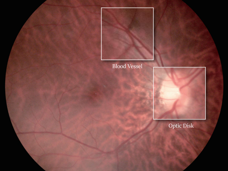
The FC161 provides high-resolution fundus images with up to 12 Megapixels. It makes the diagnosis of early lesions more accurate and is also essential for diagnosing retinal disease, such as diabetic retinopathy, glaucoma, and macular degeneration.
The FC161 provides high-resolution fundus images with up to 12 Megapixels. It makes the diagnosis of early lesions more accurate and is also essential for diagnosing retinal disease, such as diabetic retinopathy, glaucoma, and macular degeneration.
FLEXIBLE IMAGING
9 Fixation Targets
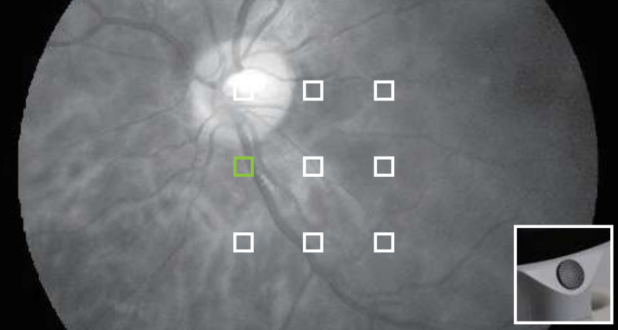
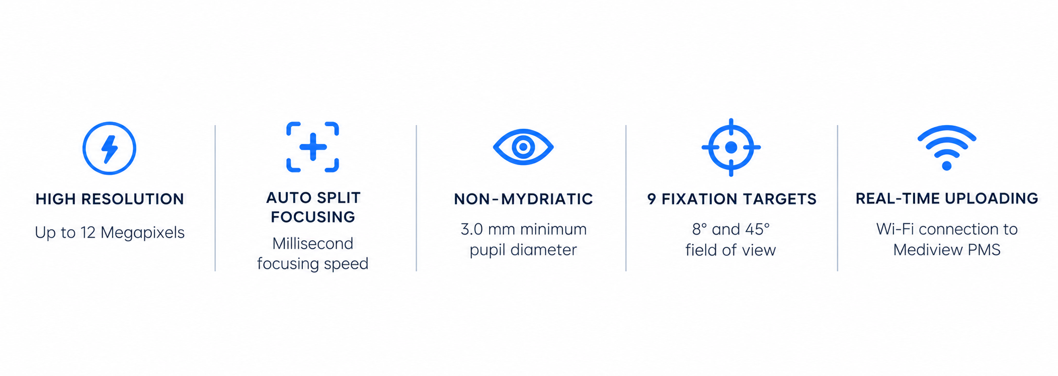
With 9 fixation targets, the FC161 covers an 8° field of view of the fundus and supports the examination of early peripheral lesions.
With a 45° field of view, a single central fundus image can be taken to meet the needs of fundus disease screening.
With 9 fixation targets, the FC161 covers an 8° field of view of the fundus and supports the examination of early peripheral lesions.
With a 45° field of view, a single central fundus image can be taken to meet the needs of fundus disease screening.
NON-MYDRIATIC
Imaging Without Dilation
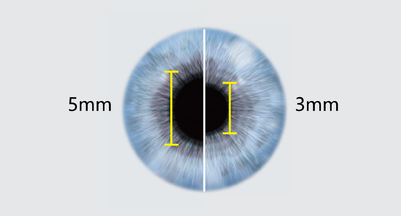
The FC161 provides high-resolution fundus images with up to 12 Megapixels. It makes the diagnosis of early lesions more accurate and is also essential for diagnosing retinal disease, such as diabetic retinopathy, glaucoma, and macular degeneration.
The FC161 provides high-resolution fundus images with up to 12 Megapixels. It makes the diagnosis of early lesions more accurate and is also essential for diagnosing retinal disease, such as diabetic retinopathy, glaucoma, and macular degeneration.
INTUITIVE DESIGN
Smart Touch Operation
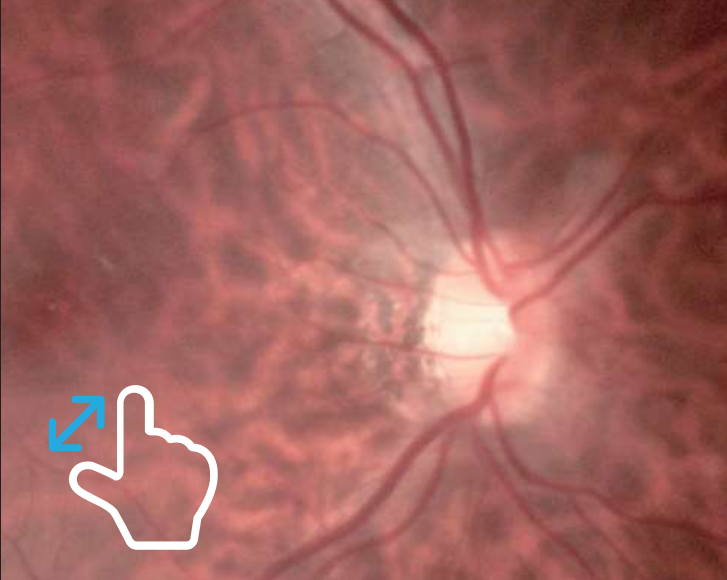
A 4.3-inch full-touch LED screen brings a smarter operating experience. Images can be zoomed at any time to check the details of the fundus and easily slide the screen to edit and view the case.
A 4.3-inch full-touch LED screen brings a smarter operating experience. Images can be zoomed at any time to check the details of the fundus and easily slide the screen to edit and view the case.
SEAMLESS WORKFLOW
Real-Time Uploading
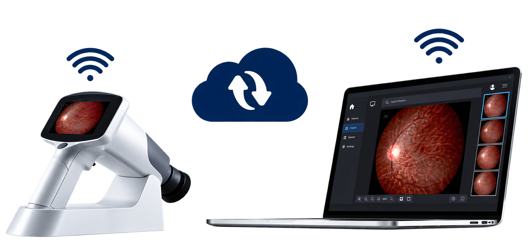
With 9 fixation targets, the FC161 covers an 8° field of view of the fundus and supports the examination of early peripheral lesions.
With a 45° field of view, a single central fundus image can be taken to meet the needs of fundus disease screening.
With 9 fixation targets, the FC161 covers an 8° field of view of the fundus and supports the examination of early peripheral lesions.
With a 45° field of view, a single central fundus image can be taken to meet the needs of fundus disease screening.

Learn more about the FC161 Hand-held Fundus Camera and how it can support retinal imaging in your practice.
Bring Retinal Imaging Into Focus
Looking for something specific?
Our team is here to help with product information, technical questions and ophthalmic imaging solutions.